
Binding-Pose Prediction
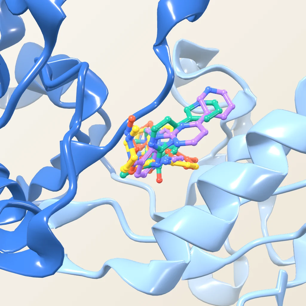
Real structure · PDB 1HSG
Generate candidate poses for a ligand in a protein's binding pocket and rank them by docking score and key contacts. A score orders poses; it is not a binding affinity.

Candidate binding poses and interface maps, ranked and explained.
 01
Target and partner are processed, with the binding site defined where applicable.
 02
Binding orientations are sampled and evaluated with the selected docking method.
 03
Representative poses are shortlisted by score, clustering, and structural assessment.
 04
Interface residues and predicted interactions of the selected complex are characterised.
Illustrative renders, not outputs from an actual run.
From predicted binding poses to structural insights for drug discovery.
Six system types, each with its own sampling and scoring considerations.
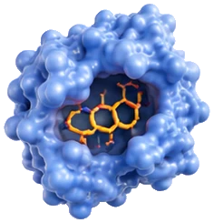
Pocket-directed docking with interaction breakdown.
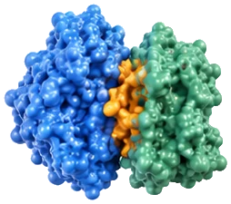
Association modes and interface characterisation.
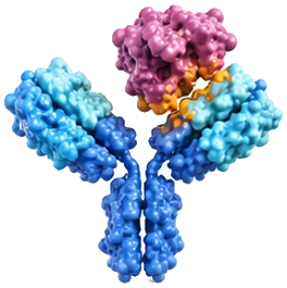
CDR-aware docking of the paratope–epitope interface.
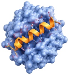
Peptide binding modes and interactions, allowing for backbone flexibility.
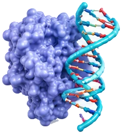
DNA recognition and binding-interface analysis, including groove contacts.
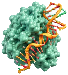
RNA interaction modes and interface mapping, allowing for RNA flexibility.
Illustrative renders of each system type.
Real structure · PDB 1HSG
Generate candidate poses for a ligand in a protein's binding pocket and rank them by docking score and key contacts. A score orders poses; it is not a binding affinity.
Real structure · PDB 1BRS
Identify where two proteins meet, mapping interface residues within 4.5 Å and listing contacts whose geometry is consistent with possible hydrogen bonds or salt bridges. These are contacts in a model, not confirmed interactions.
Real structure · PDB 3HFM
Resolve the interface pair by pair, from each contacting residue to its partner, contact type and distance in ångströms.
Illustrative data
Compare docking outcomes for the parent, a reference and AI-assisted candidates side by side under one protocol. Scores are for ordering only.
Docking proposes poses; simulation tests whether they hold. MD does not automatically validate a pose.
A high-scoring pose is a structural hypothesis, not a demonstration that two molecules bind, and docking scores are not affinities.
Explore predicted binding poses, interfaces, and residue-level interactions with a docking workflow designed around your molecular system.